Instructor Case Studies, Technology in Education
Posted on 4/19/24 by Sarah Boudreau
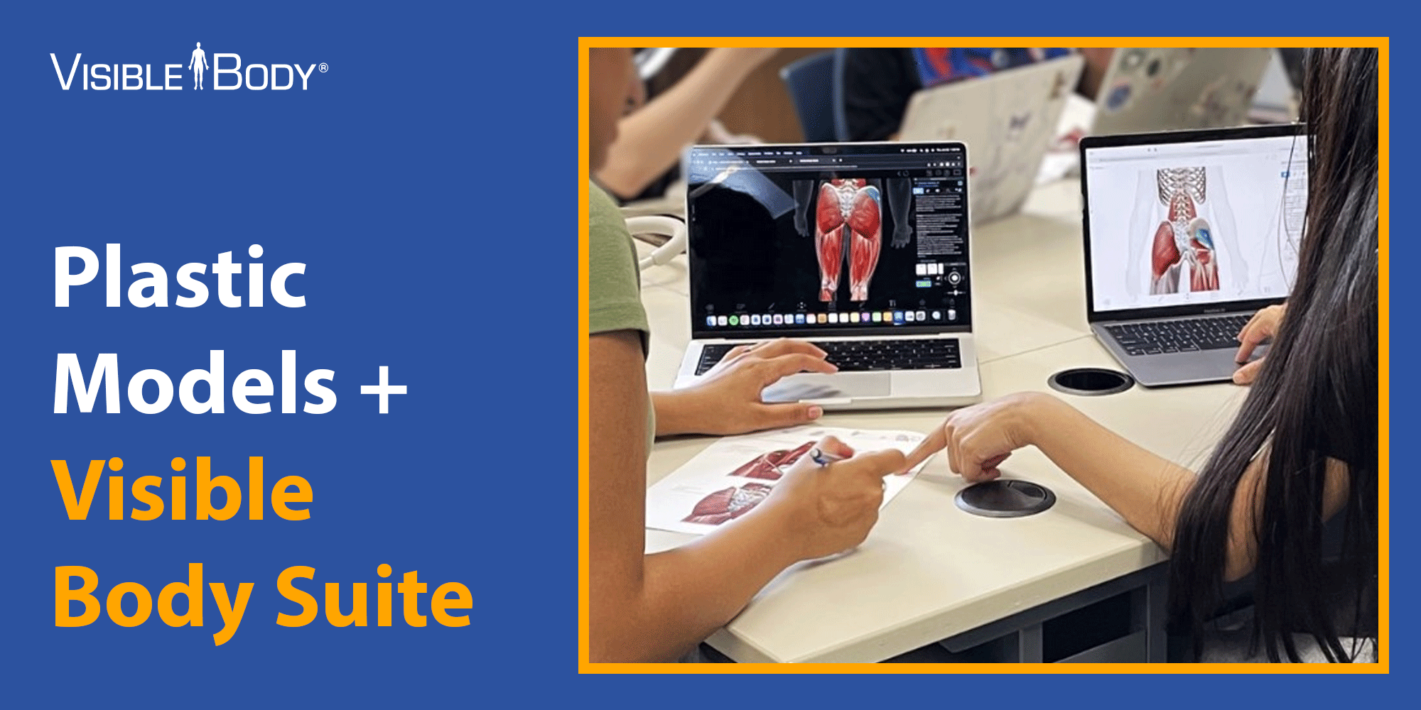
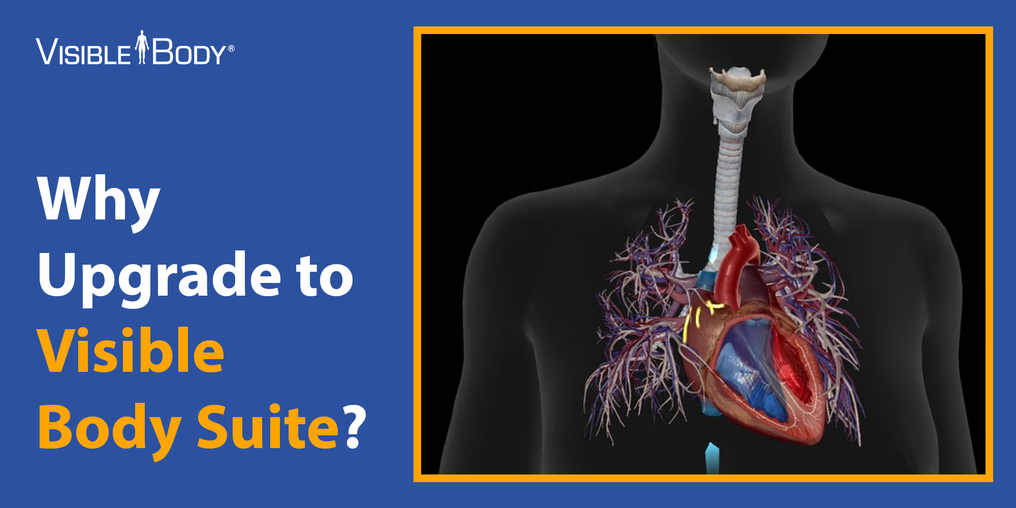
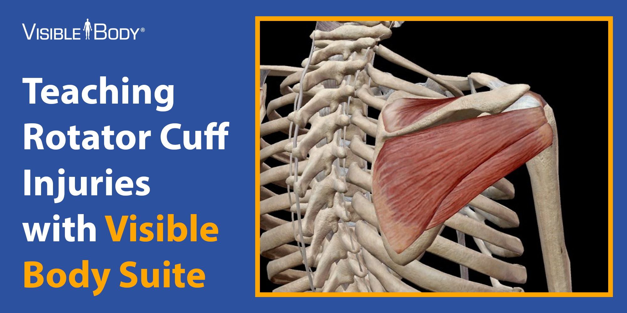
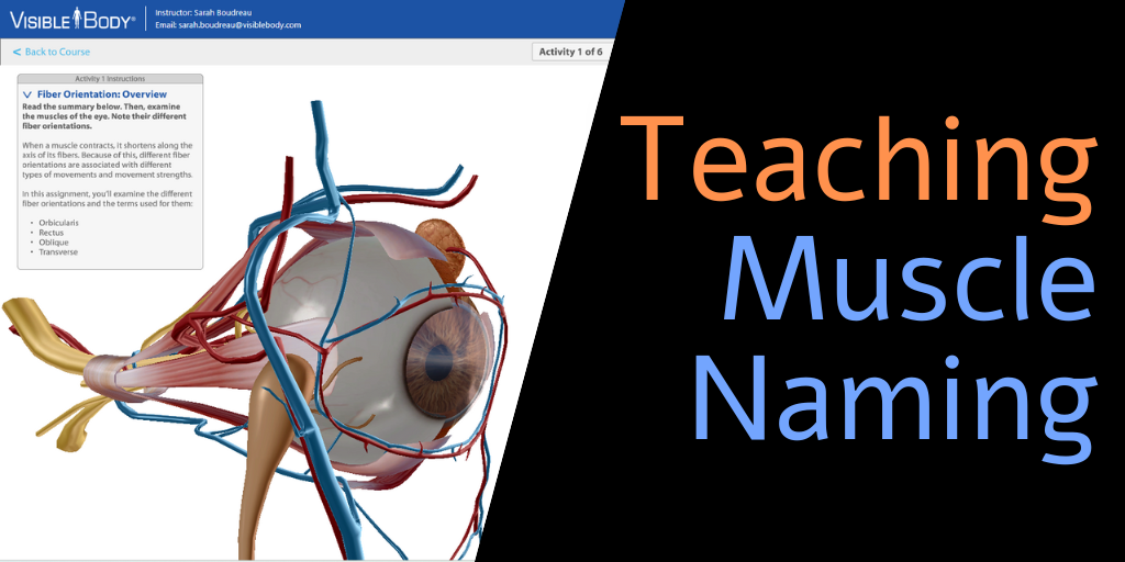
Dr. Katelyn Wood is an assistant professor in the Department of Anatomy and Cell Biology at Western University in Ontario, Canada. Dr. Wood is passionate about improving A&P instruction, and she recently presented a poster at Anatomy Connected, ...